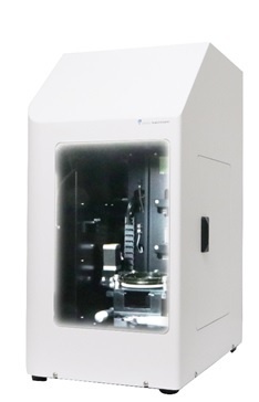
单分子检测水平的定量细胞生物学检测仪器。
受激发射损耗(STED)是一种强大的显微技术,可以观察到空间分辨率低于衍射极限的荧光结构。Alba-STED使用脉冲激发和脉冲耗尽方法(pSTED)结合数字频域荧光寿命成像(FastFLIM)来记录时间分辨光子,从而提高图像分辨率并分离具有相同激发波长的两个荧光标记物。
关键特色 : Alba-STED for FLIM/FFS:
• pSTED (Pulsed excitation and pulsed STED)
• FastFLIM for time-resolved pSTED acquisition
• Improved image resolution using the phasor plot
• Dual-label excitation
• Fast image acquisition (dwell time: 0.2 μs)
• High dynamic range (signal up to 60 million counts/s)
测试实例展示 Alba-STED for FLIM/FFS
Confocal (left) vs. pSTED (right) images of the actin labeled with the SiR dye in fixed glia cells, acquired by FastFLIM.
Confocal (left) vs. pSTED (right) images of the actin labeled with the SiR dye in fixed glia cells, acquired by FastFLIM.
Confocal images of 60nm fluorescence beads (left); pSTED images (middle); sharpening the pSTED image using a binary filter based on the phasor plots (right).
Dual labels can be separated using pSTED and FastFLIM. Atto 647N and Atto 655 were used as labels; they both are excited by the 640 nm laser. The two dyes are first separated using the phasor plots, and then assigned with two different false colors (Atto 647N - yellow, Atto 655 - purple) to produce the processed and merged pSTED image of the two labels.
参考论文:
Monomeric cohesin state revealed by live-cell single-molecule spectroscopy.
Liu, W., Biton, E., Pathania, A., Matityahu, A., Irudayaraj, J., Onn, I.
EMBO Rep. 2019 Dec 29:e48211. doi: 10.15252/embr.201948211. [Epub ahead of print]
Photon-separation to enhance the spatial resolution of pulsed STED microscopy.
Tortarolo, G., Sun, Y., Teng, K.W., Ishitsuka, Y., Lanzanó, L., Selvin, P.R., Barbieri, B., Diaspro, A., Vicidomini, G.
Nanoscale. 2019 Jan 9. doi: 10.1039/c8nr07485b. [Epub ahead of print]
A straightforward STED-background corrected fitting model for unbiased STED-FCS analyses.
Wang, R., Brustlein, S., Mailfert, S., Fabre, R., Fallet, M., Sivankutty, S., Rigneault, H., Marguet, D.
Methods. 2018 May 1;140-141:212-222. doi: 10.1016/j.ymeth.2018.02.010. Epub 2018 Feb 14.
A Novel Pulsed STED Microscopy Method Using FastFLIM and the Phasor Plots
Sun, Y.,Tortarolo G., Teng, K.-W., Ishitsuka, Y., Coskun, U.C., Liao, S.-C., Diaspro, A., Vicidomini, G., Selvin, P.R., Barbieri, B.
Proc. SPIE 10069, Multiphoton Microscopy in the Biomedical Sciences XVII, 100691C (February 21, 2017)